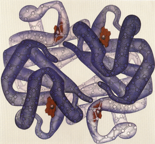
Hemoglobin
1978, Unknown Dimensions

Geis illustrates the hemoglobin molecule as four symmetrically arranged myoglobins. Since it is responsible to the transport of oxygen, it can change from an oxygen-binding configuration to an oxygen-releasing configuration in response to the demand for oxygen.
Used with permission from the Howard Hughes Medical Institute (www.hhmi.org). All rights reserved.

Related PDB Entry: 2DHB
Experimental Structure Citation
Bolton, W. & Perutz, M. F. (1970). Three dimensional Fourier synthesis of horse deoxyhaemoglobin at 2.8 Å resolution. Nature, 228, 551-552.
About Hemoglobin
Molecule of the Month: Hemoglobin
Hemoglobin is a protein found in the red blood cells of all vertebrates that is responsible for the transport of oxygen. The high iron concentration in the molecule gives blood its red color. Hemoglobin has a tetrameric quaternary structure of two alpha and two beta chains, each containing a ring-shaped heme group, giving the overall appearance of having four myoglobins combined into one structure. These heme groups use their central iron atom to bind oxygen, and, in this way, blood carries oxygen from the respiratory organs throughout the body. While hemoglobin is best known for its role in vertebrate respiration, it is also found in some invertebrates, fungi, and plants, where it transports other gases like carbon monoxide, nitric oxide, and hydrogen sulfide.
Text References
Dutta, S. & Goodsell, D. (2003). Molecule of the Month: Hemoglobin. DOI: 10.2210/rcsb_pdb/mom_2003_5
Initial Structure Determination Reference
Perutz, M. F., Rossmann, M. G., Cullis, A. F., Muirhead, H., & Will, G. (1960). Structure of hæmoglobin: a three-dimensional Fourier synthesis at 5.5-Å. resolution, obtained by X-ray analysis. Nature, 185, 416-422.


