3D Print: Alpha-amylase
Download curated files for 3D printing.
3D Model Information
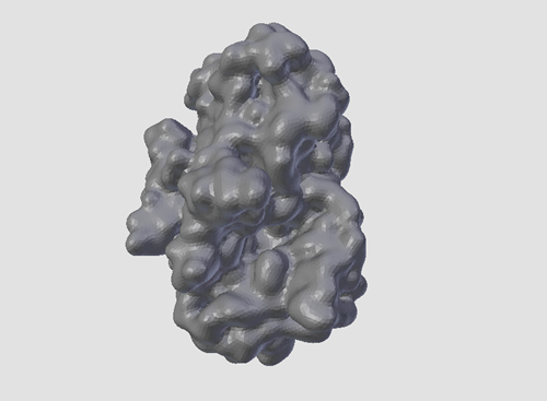
PDB structure used to generate the model file: 1ppi
Model generated with: Molecular Maya/Autodesk Maya
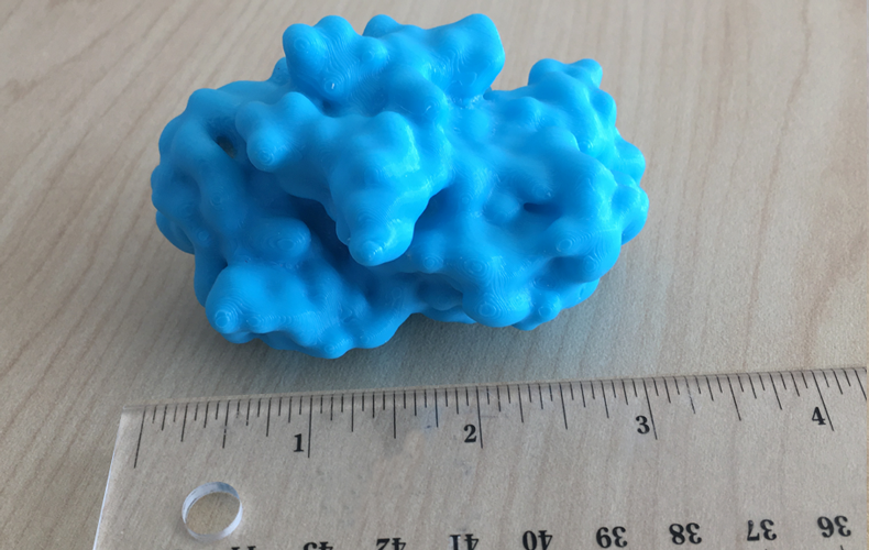
Model physical size (in mm): 53.9 x 81.4 x 61.8
Download 3D Print File (.stl)Sample print
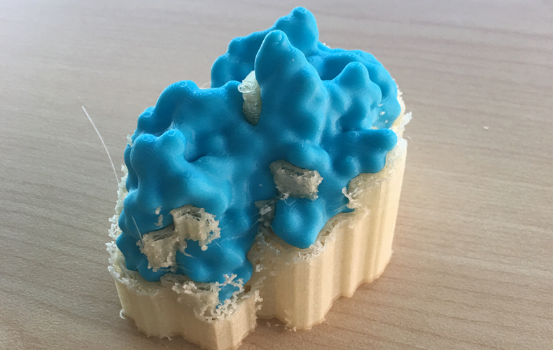
Printed on: Ultimaker S3
Model material: PLA
Support material: PVA (water soluble)
Output scale: 100%
3D Print and Structural Features of Alpha-amylase
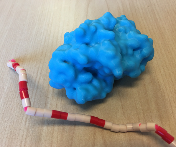
Image 1
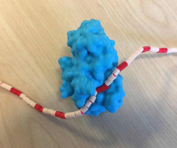
Image 2
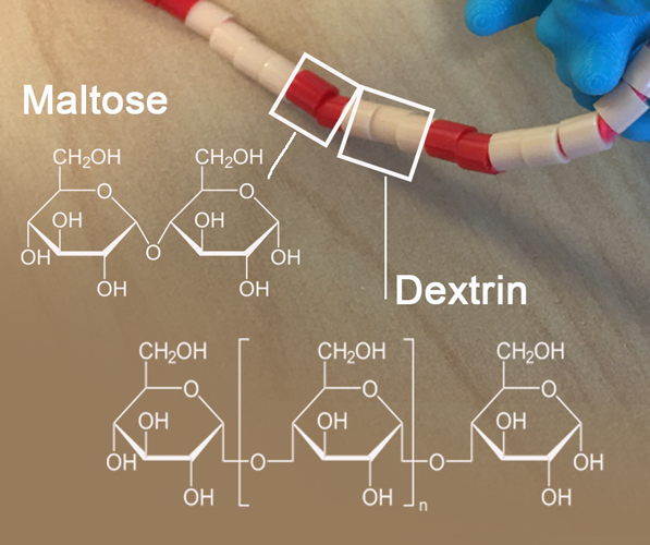
Image 3
Alpha-amylase is a small enzyme found in saliva (produced by the salivary glands), and small intestine (produced in the pancreas). The enzyme starts the digestion of starches by cleaving the long carbohydrate chains into maltose and dextrin.
This printed model can be used to demonstrate how the substrate interacts with the enzyme’s active site (Image 1).
The carbohydrate chain was modeled by threading 5mm melty beads onto a pipe cleaner and then inserted into the groove that represents the active site (Image 2).
Each bead represents one glucose. 2 colors were used to indicate the possible products of multiple reactions of the enzyme with the chain (past cleavage and exiting the active site): red for maltose, white for dextrin (Image 3).
Related PDB-101 Learning Resources
More about Alpha-amylase
Browse Enzymes
Browse Biotechnology
Browse Biological Energy
