2. TBP-DNA
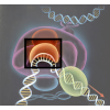
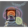
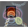
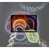
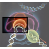
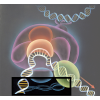
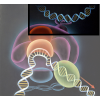
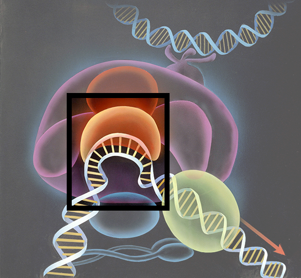
Geis depicts the TATA-Binding Protein sitting on top of the DNA curvature, matching the description of a molecular “saddle.” The DNA strand, colored with a blue backbone and yellow ladder bases, is seen partially unwound where TBP has bound.

Used with permission from the Howard Hughes Medical Institute (www.hhmi.org). All rights reserved.

Related PDB Entry: 1VTL
Experimental Structure Citation
Kim, J. L., Nikolov, D. B., & Burley, S. K. (1993). Co-Crystal Structure of TBP Recognizing the Minor Groove of a TATA Element. Nature, 365, 520-527.
About TBP-DNA
TBP is able to recognize the special TATA sequence. The binding of TBP causes DNA to partially unwind and creates two sharp kinks on either side of the sequence so DNA is bent. When bound, TBP resembles a molecular “saddle” sitting on the DNA strand.
Text References
Kim, J. L. & Burley, S. K. (1994). 1.9 Å resolution refined structure of TBP recognizing the minor groove of TATAAAAG. Nat Struct Biol. 1, 638-653.
