Molecular Landscapes by David S. Goodsell
Cellulose Synthase, 2021

Acknowledgement: Illustration by David S. Goodsell, RCSB Protein Data Bank. doi: 10.2210/rcsb_pdb/goodsell-gallery-029
In this cross-section, a plant cell is shown at the bottom, with the plasma membrane in green and the cell cytoplasm in blue, with a microtubule running horizontally just under the membrane. Two cellulose synthase complexes are building cellulose fibers (tan) outside the cell. The top portion of the illustration is filled with molecules of the cell wall, including xyloglucans and pectins in darker brown filling in the spaces between the cellulose fibers.
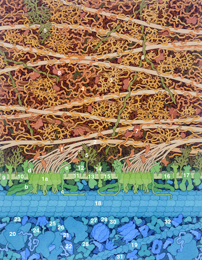
Cellulose Synthase Complex
Cellulose synthase lays down cellulose fibers in specific orientations by moving along microtubules. Many proteins form a complex with cellulose synthase and assist with the process. The details of this complex are speculative and are based on the sizes and structures (if known) of the component proteins.
1a. Cellulose synthase
b. Cellulose synthase interactive 1 (CSI1)
c. Companion of cellulose synthase (CC)
d. Endoglucanase 25 (KOR)
e. Protein COBRA (COB)
f. Chitinase-like protein 1 (CTL1)
Cell Wall
The cell wall is composed of a complex network of fibrous molecules that are composed of carbohydrates and proteins:
2. Cellulose fibrils are thought to be composed of 18 cellulose chains; each cellulose chain is a linear strand of glucose units
3. Pectins are long, branched polysaccharides
4. Xyloglucans are long polysaccharides with many small branches
5. Arabinogalactan proteins are unstructured protein chains decorated with branched carbohydrates; some have attached lipids and are bound to the plasma membrane
6. Extensin is a protein that forms networks that strengthen the cell wall
7. Expansin helps loosen the interactions of carbohydrates
8. Cell wall invertase
Plasma Membrane
The plasma membrane contains many proteins involved in transport and sensing. The ones shown here include:
9. H+ ATPase 1 (AHA1)
10. Inorganic phosphate transporter
11. Sugar transport protein 1 (STP1)
12. Fasciclin-like arabinogalactan protein 4 (FLA4, SOS5)
13. Calcium-transporting ATPase 8 (ACA8)
14. LRR receptor-like serine/threonine-protein kinase FEI 1
15. Sucrose transport protein SUC3
16. Aquaporin PIP2-1
17. ABC transporter ABCB.1
Cytoplasm
The cytoplasm is filled with molecules involved in protein synthesis, cell metabolism, and the cytoskeleton. These include:
18. Microtubule
19. Actin filament
20. Ribosome
21. Transfer RNA and elongation factor
22. Aminoacyl-tRNA synthetase
23. UTP:glucose-1-phosphate uridylyltransferase 1 (UGP1)
24. UDP-glucose 6-dehydrogenase 1 (UGD1)
25. Trifunctional rhamnose biosynthetic enzyme 1 (RHM1)
26. Hexokinase 1 (HXK1)
27. Phosphoglycerate mutase 1 (PGM1)
28. Sucrose synthase 1 (SUS1)
29. Fructokinase 1
30. Cytosolic invertase 1 (CINV1)
31. Cytosolic glucose-6-phosphate isomerase (PGIC)
32. UDP-glucose 4-epimerase 2 (UGE2)
Note that many of the structures linked here are from related organisms.
Select References
- Lampugnani ER, Khan GA, Somssich M, Persson S (2018) Building a plant cell wall at a glance. J. Cell Sci. 131, jcs207373.
- Hofte H, Voxeu A (2017) Primer: Plant cell walls. Curr. Biol. 27, R853-R909.
- Li S, Bashline L, Zheng Y, Xin X, Huang S, Kong Z, Kim SH, Cosgrove DJ, Gu Y (2016) Cellulose synthase complexes act in a concerted fashion to synthesize highly aggregated cellulose in secondary cell walls of plants. PNAS 113, 11348-11353.
- Cosgrove DJ (2014) Re-constructing our models of cellulose and primary cell wall assembly. Curr. Opin. Plant Biol. 22, 122-133.
- Dunkley TPJ, Hester S, Shadforth IP, Runions J, Weimar T, Hanton SL, Griffin JL, Bessant C, Brandizzi F, Hawes C, Watson RB, Dupree P, Lilley KS. (2006) Mapping the Arabidopsis organelle proteome. PNAS 103, 6518-6523.
- Somerville C, Bauer S, Brininstool G, Facette , Hamann T, Milne J, Osborne E, Paredez A, Persson S, Raab T, Vorwerk S, Youngs H (2004) Towards a systems approach to understanding plant cell walls. Science 306, 2206-2211.
- Carpita NC, Gibeaut DM (1993) Structural models of primary cell walls in flowering plants: consistency of molecular structure with the physical properties of the walls during growth. Plant J. 3, 1-30.
