Molecular Landscapes by David S. Goodsell
SARS-CoV-2 Fusion, 2020

Acknowledgement: Illustration by David S. Goodsell, RCSB Protein Data Bank; doi: 10.2210/rcsb_pdb/goodsell-gallery-026
This painting depicts the fusion of SARS-CoV-2 (magenta) with an endosomal membrane (green), releasing the viral RNA genome into the cell cytoplasm (blue), where it is beginning to be translated by cellular ribosomes to create viral polyproteins. The painting includes speculative elements that are designed to highlight the process, most notably, multiple states of the viral spike protein are shown.
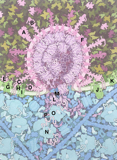
A. Pre-fusion state of the viral spike protein (6crz)
B. Viral spike protein S2 domain, after S1 is released.
C. Viral spike protein inserting into the endosomal membrane
D. Post-fusion state of the viral spike protein (6xra)
E. S1 domain of viral spike
F. Complex of viral M, E (5x29), ORF3a (6xdc) and ORF7a (6w37)
G. ACE2 (6m17)
H. LAMP (5gv0)
I. ABC transporter
J. V-ATPase (5vox)
K. Mucolipin (5wj5)
L. Viral nucleocapsid protein (6m3m, 6wzo)
M. Viral RNA genome
N. Ribosomal initiation complex
O. Translating ribosome
P. Nascent viral polyprotein
This painting was created as part of the show "New Ways of Living: Understanding the Science of COVID-19," in association with SciCommMake 2020.
Selected References
Ke, Z., et al. (2020) Structures and distributions of SARS-CoV-2 spike proteins on intact virions. Nature https://doi.org/10.1038/s41586-020-2665-2
Yao, H., et al. (2020) Molecular architecture of the SARS-CoV-2 virus. Cell 183, 730-738.
Zeng, W., et al. (2020) Biochemical characterization of SARS-CoV-2 nucleocapsid protein. Biochem. Biophys. Res. Comm. 527, 618-623.
Li, F. (2016) Structure, function, and evolution of coronavirus spike proteins. Annu. Rev. Virol. 3, 237-261.
Liu, D.X, et al. (2014) Accessory proteins of SARS-CoV and other coronaviruses. Antiviral Res. 109, 97-109.
Sonenberg, N. and Hinnebusch, A. G. (2009) Regulation of translation initiation in eukaryotes: mechanisms and biological targets. Cell 136, 731-745.
