Folding of Protein Domains
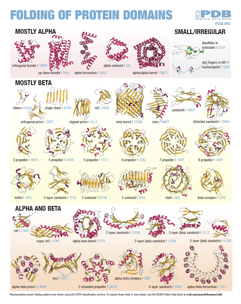
Proteins are polymers of amino acids. The secondary structure elements are beta sheets, alpha helices and loops. These elements are arranged in 3D space creating the environment for the protein or protein domain to fulfill its function.
As more and more 3D shapes became known, researchers noticed patterns in the folding of whole proteins or protein domains.
This poster highlights common arrangements of secondary structure elements in protein domains as defined using the CATH classification scheme.
Exploring the Protein Domains in 3D
Use the PDB IDs provided on the poster to explore these domains on RCSB.org in 3D using MolStar (Mol*). Enter the PDB ID in the Import > Download Structure section in the viewer's right menu.
You can take the exploration a step further and create paper models of these domains. The activity offers three domain building instructions as examples.

