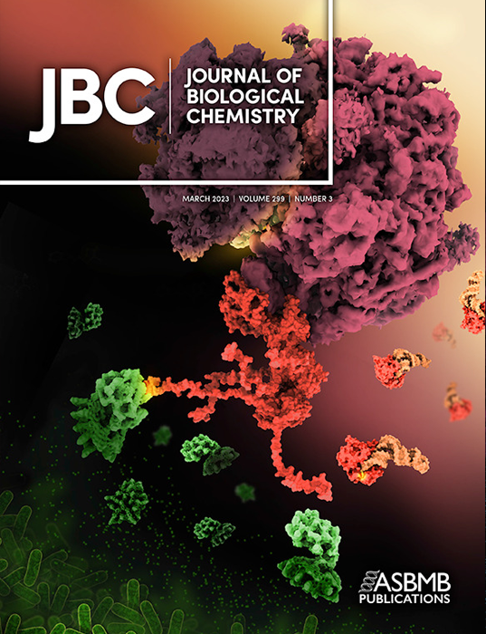
Shiga Toxin 2 in Complex with Ribosomal P-stalk

Some of bacterial ribosome-inactivating toxins evolved to mimic the mechanism employed for loading onto the ribosome by translation factors. The illustration depicts how Shiga toxin 2a (Stx2a) holotoxin (green, PDB ID: 7U6V, EMD-26381) outcompetes the EF-Tu elongation factor (red, PDB ID: 1TTT) complexed with Phe-tRNAPhe (orange) for binding to the ribosomal P-stalk (red) at the large subunit of the 80S ribosome (purple, EMD-26380). Kulczyk et al. determined a cryo-EM structure of Stx2a bound to the native ribosomal P-stalk. The illustration was created by Maria Voigt (RCSB PDB) with materials provided by Arek Kulczyk (Rutgers University, Institute for Quantitative Biomedicine).
This image appeared on the March 2023 cover of the Journal of Biological Chemistry.
Cryo-EM structure of Shiga toxin 2 in complex with the native ribosomal P-stalk reveals residues involved in the binding interaction (2023)
AW Kulczyk, COS Sorzano, P Grela, M Tchórzewski, NE Tumer, XP Li
J Biol Chem 299:102795.
https://doi.org/10.1016/j.jbc.2022.102795
