G Protein-Coupled Receptor (GPCR) Paper Model
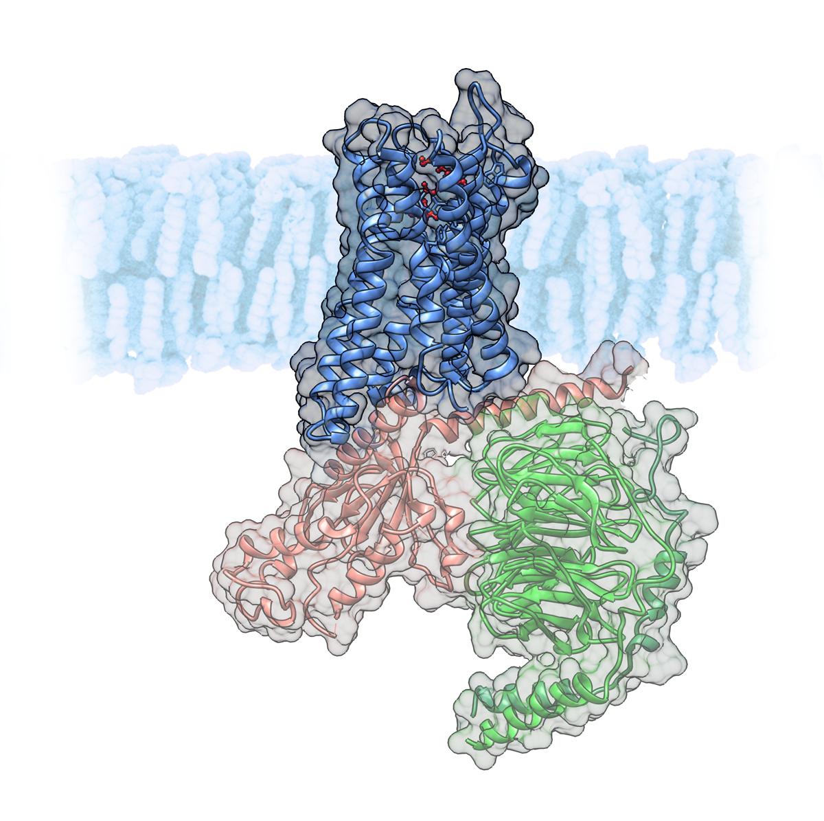
On the extracellular side, the helices form a cavity where ligands (e.g. endorphins, morphine, serotonin) bind. On the intracellular side, the receptor is coupled to G protein. When the receptor is activated by a ligand, the G protein splits in two parts which then activate other proteins in the internal signal transduction pathways.
Examples of GPCRs are opioid receptors, rhodopsin or adrenergic receptors.
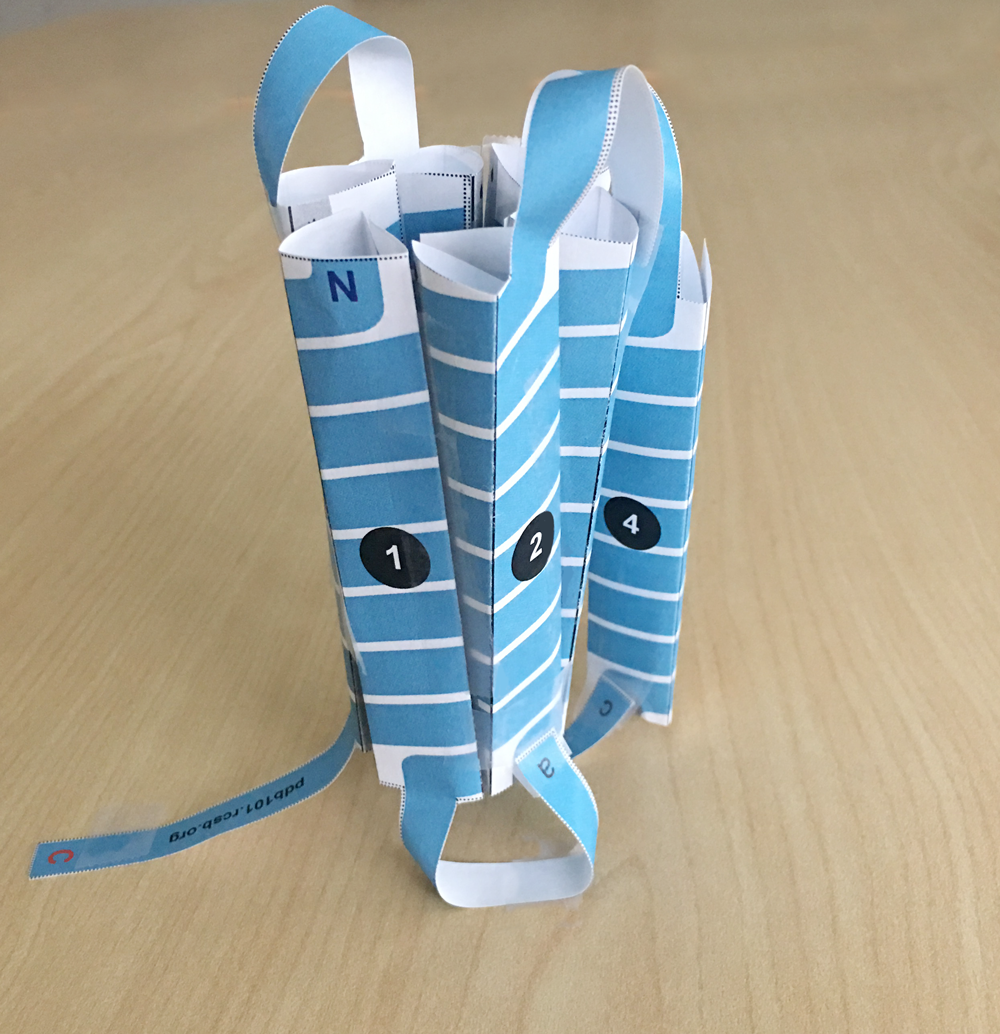
To build the paper model featuring the shared structural features of all GPCRs download and print the template PDF.
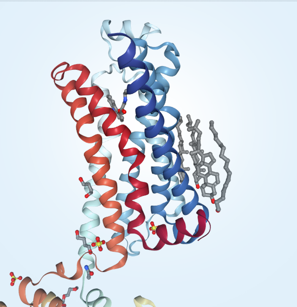
This paper model is not based on any particular PDB structure. Explore examples of GPCRs in 3D: 6dde (opioid receptor), 2rh1 (adrenergic receptor), and 1f88 (rhodopsin).
Video Instructions: How to fold the GPCR paper model
Use the PDB-101 Browser to explore more resources and articles related to Drugs and the Brain .
