This exercise is a part of RCSB PDB Curriculum Module Biomolecular Structures and Models and was developed as part of the RCSB Collaborative Curriculum Development Program 2015.
Exploring a Protein Structure in the RCSB PDB: Insulin
Learning Goals:
- Visualize the structure of a given molecule using RCSB PDB resources.
- Explore the structure to understand its structure function relationships
Exercise:
Review the Molecule of the Month feature on Insulin for background information. What are the main ideas of this feature?Note that there are a few PDB entries listed throughout the feature. For example, PDB entry 4ins can be linked from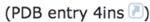
Open the Structure Summary page for the PDB entry 4ins (http://www.rcsb.org/pdb/explore/explore.do?structureId=4ins).
Read/review the page and answer the following questions based on the descriptions provided:
What is the source (organism) of the insulin molecule in this structure?
Name the authors who solved the structure of this protein?
Explore the 3-D structure of this protein by clicking on JSmol (hyperlink) under the image as seen below:
Change the viewer to PV
- Go back to the JSmol view of the PDB entry.
Are these S-S bonds within the same polymer chain or between different chains of insulin? (Hint: Color the ribbons by sequence to see if the S-S bonding is between the same or different polymer chains).
What do you think is the role of these S-S bonds? Describe in 1-2 sentences.

View the polymer chains shown to contain helical ribbons (in magenta), arrows (in golden yellow) and coil-like regions (white/grey).
Mouse-over the small grey atom (highlighted by a red arrow, in the figure below). What is it and why do you see this atom in the insulin structure (Hint: read the title and abstract of the structure for clues)


The default view is colored by chain (i.e. each protein (polymer) chain in the structure is colored in a different color.
Based on the 3-D model that you see here describe the overall composition of insulin – how many and what chains are present in the structure. Also describe the structure of each insulin molecule in terms of the helical, arrow-like or coiled regions in each chain.
In the various options click on the box next to S-S bonds to show the disulfide bonds in the structure.

Note yellow SS bond lines appear in the model. These bonds are formed by oxidation of two specific sulfur-containing amino acids. How many such bonds do you see?
