Molecule of the Month: Enoyl-CoA Carboxylases/Reductases
Enzymes that can quickly and efficiently fix carbon
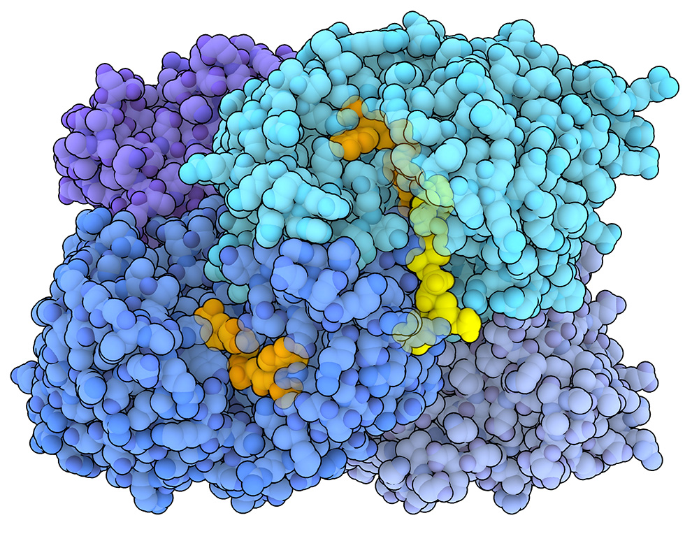
The enoyl-CoA carboxylases/reductases from Kitasatospora setae (PDB 6OWE) is a tetramer. NADPH, shown in orange, acts as an electron donor, while the substrate, an enoyl-CoA molecule, is shown in yellow.
Download high quality TIFF image
Carbon fixation is a crucial biological process that transforms carbon dioxide (CO₂), a highly abundant but biologically inaccessible molecule, into organic compounds such as sugars. Most carbon fixation is carried out by plants and algae through the enzyme Rubisco. Rubisco, however, is a relatively slow and inefficient enzyme. This inefficiency has driven evolutionary adaptations in algae and plants, such as producing large amounts of Rubisco and storing it in specialized organelles (e.g. pyrenoids and chloroplasts). Rubisco inefficiency is, in large part, due to affinity for both CO₂ and molecular oxygen (O₂), both of which are present at high concentrations in the earth's atmosphere. Despite various laboratory attempts to enhance Rubisco's efficiency, significant improvements remain elusive. Carbon fixation, however, is not exclusive to plants and algae. Certain bacteria and archaea have evolved alternative carbon fixation pathways that do not rely on Rubisco. Studying these systems can provide insights into how we might engineer synthetic systems to capture and fix carbon dioxide from our atmosphere, an important step towards reducing atmospheric CO₂.
A more efficient way of fixing carbon
A relatively recently discovered family of enzymes known as enoyl-CoA carboxylases/reductases (ECRs), which can be found in some species of bacteria and archae, have been found to catalyze carbon fixation at exceptionally fast rates. An ECR from a soil bacterium called Kitasatospora setae (PDB 6OWE) is shown on the right. Unlike Rubisco, ECRs do not accept O₂ as a substrate, thus avoiding possible competition with CO₂. Remarkably, ECRs have been found to outperform Rubisco by up to 10-fold in terms of reaction rate. Recent structural studies have shown that ECRs function as homotetramers, wherein each subunit can bind to CO₂, NADPH (which acts as a cofactor and an electron donor), and a substrate molecule (an enoyl-CoA). During carbon fixation, a hydride ion (H-) is transferred from NADPH to the substrate molecule. This step makes the substrate highly reactive, allowing it to react with the bound carbon dioxide to form a new product (an alkyl-coA ester).
Enzymatic Synchronization
What accounts for the impressive speed and efficiency of ECRs in carbon fixation? Recent studies have uncovered a unique synchronization mechanism that facilitates rapid catalysis. The tetrameric ECR adopts a "pair of dimers" configuration, wherein each subunit forms a dimer with a neighboring subunit, and the two dimers assemble into an "X"-shaped geometry. This special arrangement allows for coordinated action during the catalytic cycle, which is shown in the animation on the left. In the initial empty state (PDB 6NA3), the enzyme is symmetric, but upon binding to NADPH (shown in orange), the tetramer undergoes a conformational change (PDB 6NA6). One subunit of each dimer closes in a coordinated manner, precisely positioning the substrate (shown in yellow), NADPH, and carbon dioxide (which is not shown in the animation) in close proximity for carbon fixation (PDB 6NA4). Once the reaction is complete, the subunits reopen to release the product. This release event triggers the partner subunit within the dimer to close, initiating the next round of catalysis. Synchronized switching between open and closed states is coupled with a twisting motion of the enzyme complex, which is thought to enhance substrate binding and/or product release. The rapid and coupled synchronization of the ECR's subunits is believed to be a key factor contributing to superior performance versus Rubisco.
Exploring the Structure
Taking a closer look at carbon fixation
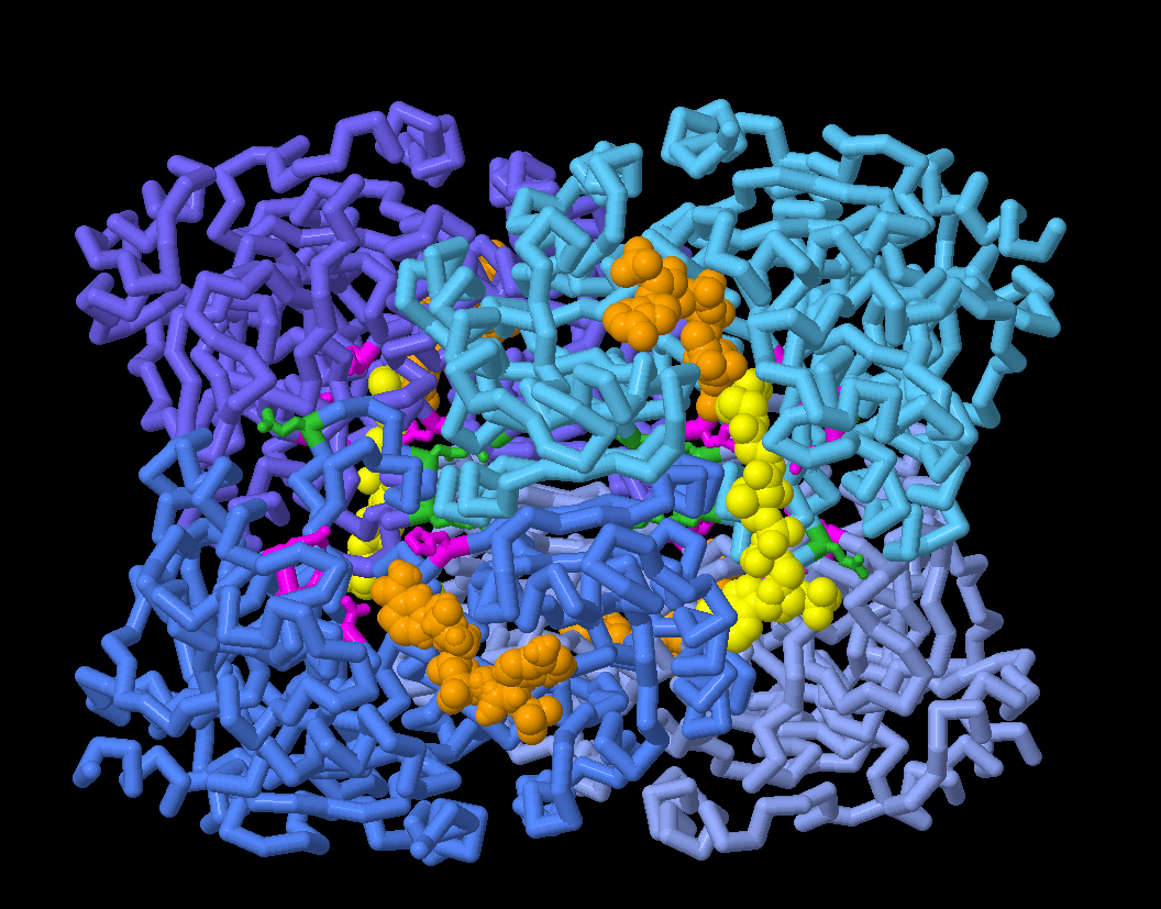
In the JSmol tab, you can explore the structures of different ECR conformations and take a closer look at amino acid residues thought to be involved in binding carbon dioxide (shown in magenta), plus residues involved in coordinating enzymatic synchronization (shown in green).
Topics for Further Discussion
- Rubisco, considered to be one of the most abundant enzymes on the planet, is used by plants and algae to fix carbon dioxide.
- Cyanobacteria also use Rubisco, storing multiple copies of the enzyme in an organelle called the carboxysome. Read more about this and other carbon capture mechanisms
- This study took advantage of time-resolved crystallography techniques, which have also been used to study other enzymes that rapidly catalyze reactions, including studies of light activatable proteins.
Related PDB-101 Resources
References
- 6OWE: Stoffel GMM, Saez DA, DeMirci H, Vögeli B, Rao Y, Zarzycki J, Yoshikuni Y, Wakatsuki S, Vöhringer-Martinez E, Erb TJ. (2019) Four amino acids define the CO2 binding pocket of enoyl-CoA carboxylases/reductases. Proc Natl Acad Sci U S A. 2019 Jul 9;116(28):13964-13969.
- 6NA3, 6NA4, 6NA5, 6NA6: DeMirci H, Rao Y, Stoffel GM, Vögeli B, Schell K, Gomez A, Batyuk A, Gati C, Sierra RG, Hunter MS, Dao EH, Ciftci HI, Hayes B, Poitevin F, Li PN, Kaur M, Tono K, Saez DA, Deutsch S, Yoshikuni Y, Grubmüller H, Erb TJ, Vöhringer-Martinez E, Wakatsuki S. (2022) Intersubunit Coupling Enables Fast CO2-Fixation by Reductive Carboxylases. ACS Cent Sci. 2022 Aug 24;8(8):1091-1101.
- Schwander T, Schada von Borzyskowski L, Burgener S, Cortina NS, Erb TJ.(2016) A synthetic pathway for the fixation of carbon dioxide in vitro. Science. Nov 18;354(6314):900-904.
March 2025, Janet Iwasa
http://doi.org/10.2210/rcsb_pdb/mom_2025_3