Biomolecular Structural Biology
methods for determining atomic structures
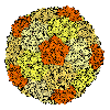
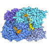
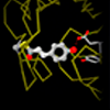
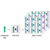
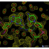
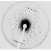
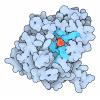
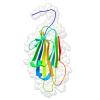
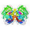
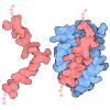
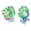
Scientists use a variety of experimental methods to discover the inner workings of biological molecules. These include X-ray crystallography, NMR spectroscopy, and electron microscopy. Each method has specific advantages for the exploration of biological molecules.
Molecule of the Month Articles (12)
![]() | Adenine Riboswitch in Action XFEL serial crystallography reveals what happens when adenine binds to a riboswitch |
![]() | Fifty Years of Open Access to PDB Structures The Protein Data Bank is celebrating its golden anniversary! |
![]() | Lysozyme Lysozyme attacks the cell walls of bacteria |
![]() | Myoglobin Myoglobin was the first protein to have its atomic structure determined, revealing how it stores oxygen in muscle cells. |
![]() | Nanodiscs and HDL Nanodiscs conveniently package a small piece of membrane for experimental studies. |
![]() | PDB Pioneers A dozen historic structures set the foundation for the PDB archive |
![]() | Pepsin Pepsin digests proteins in strong stomach acid |
![]() | Photoactive Yellow Protein Researchers use synchrotrons and X-ray lasers to reveal the rapid processes of light sensing. |
![]() | Ribonuclease A Ribonuclease cuts and controls RNA |
![]() | Selenocysteine Synthase Selenium is used in place of sulfur to build proteins for special tasks |
![]() | Trypsin An activated serine amino acid in trypsin cleaves protein chains |
![]() | Twenty Years of Molecules Celebrating the structural biology revolution |
Learning Resources (13)
![]() | Quasisymmetry in Icosahedral Viruses Activity Page Build 3D paper models of several viruses to explore how quasisymmetry builds capsids with different sizes. |
![]() | Expanding Boundaries of Complexity with 3DEM Flyer 3D electron microscopy (3DEM) is revolutionizing the field of structural biology. |
![]() | 200 Icosahedral Viruses Poster |
![]() | Photoactive Yellow Protein and XFEL/SFX GIF Structures of photoactive yellow protein were determined by serial femtosecond crystallography after illumination, capturing the isomerization of the chromophore after it absorbs light. Structures included in this movie include: 5hd3 (ground state), 5hdc (100-400 femtoseconds after illumination), 5hdd (800-1200 femtoseconds), 5hds (3 picoseconds), 4b9o (100 picoseconds), 5hd5 (200 nanoseconds) and 1ts0 (1 millisecond). For more, see the Molecule of the Month on Photoactive Yellow Protein and Guide to Understanding PDB Data: Methods for Determining Atomic Structures |
![]() | Celebrating 50 Years of the Protein Data Bank Archive Video In 1971, the structural biology community established the single worldwide archive for macromolecular structure data–the Protein Data Bank (PDB). From its inception, the PDB has embraced a culture of open access, leading to its widespread use by the research community. PDB data are used by hundreds of data resources and millions of users exploring fundamental biology, energy, and biomedicine.
This video looks at the history and the milestones that shaped the PDB into the leading resource for research and education it is today. |
![]() | Discovering Biology Through Crystallography Coloring Book Color the diverse 3D shapes studied by crystallographers. Created with support from the ACA. Available as a PDF and individual images. |
![]() | Biological Assemblies Guide |
![]() | Methods for Determining Structure Guide |
![]() | Resolution Guide |
![]() | R-value and R-free Guide |
![]() | Structure Factors and Electron Density Guide |
![]() | Molecular Backgrounds For Virtual Meetings Other Resource Download images created by David Goodsell to add a molecular backdrop to your next virtual meeting. Click on the image to expand. |
![]() | PDB50 the Game Other Resource A PDB “worker placement” board game that explores the process of structure discovery
|
Curriculum Resources (11)
Structural Biology Highlights (6)
Geis Digital Archive (6)
![]() | Myoglobin Geis highlights the hundreds of chemical bonds in the lattice of myoglobin. |
![]() | Myoglobin Fold Geis illustrated the structure of myoglobin, focusing on the folding pattern of the secondary structure of the protein. Unlike previous myoglobin Illustrations, this painting focuses on the tertiary structure of the molecule rather than the sequence or surface. |
![]() | Ribonuclease S Geis illustrates the structure of the ribonuclease S that highlights the dinucleotide RNA substrate in red and the four disulfide bonds in yellow. |
![]() | Trypsin Geis illustrates the structure of bovine trypsin, an enzyme that breaks down proteins, which was first revealed by X-ray crystallography in 1971 and further explored in 1974 (Krieger et al., 1974). This illustration was originally published in Scientific American (Stroud, 1984). Trypsin is a protease, an enzyme that catalyzes cleavage of polypeptide chains (Stroud, 1984). Geis' sketch depicts the structure with a ball-and-stick model and displays the sidechains of aspartic acid (Asp102), histidine (His57), and serine (Ser195), known as the catalytic triad. |
![]() | Crambin In Hendrickson and Teeter's molecular study in 1981, the crystal structure of crambin, a small seed storage protein, was determined based on the location of sulfur atoms in the protein. Using an artistic approach, Geis utilizes bright yellow shading and orange coloring to highlight the importance of these 6 sulfur atoms in this ball-and-stick representation. The backbone of the protein is depicted in blue. |
![]() | Myohemerythrin The colored print depicts the structure of myohemerythrin, which was first revealed by X-ray crystallography in 1975 (Hendrickson et al., 1975) and further refined in 1987 (Sheriff et al., 1987). Geis's illustration depicts the tertiary structure of the protein, highlighting the four anti-parallel alpha-helices and the presence of mu-oxo-diiron (iron atoms in red and oxygen atom in pink) located within the core of the macromolecule (Myohemerythrin). |