New Calendar Celebrates 50 Years of the PDB
12/31
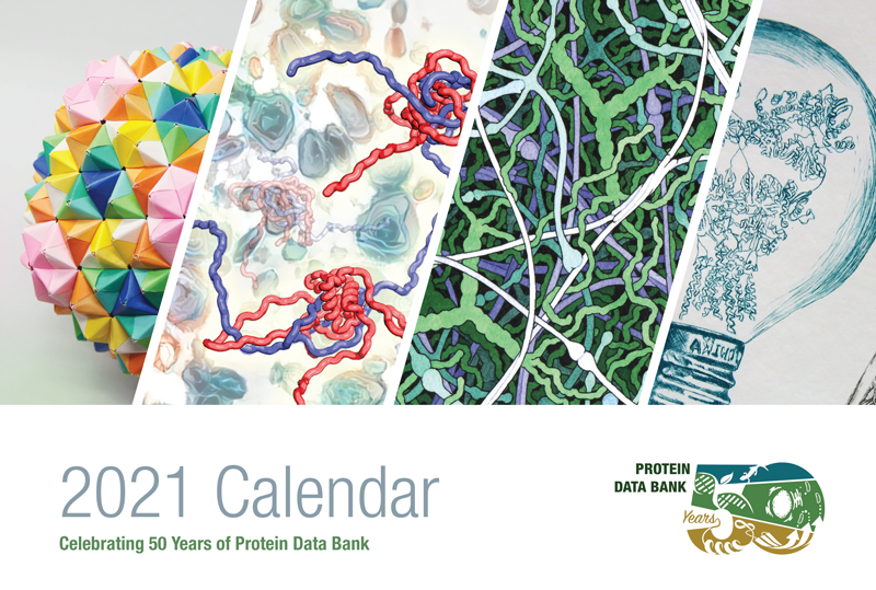
The PDB was announced on October 20, 1971 in Crystallography: Protein Data Bank Nature New Biology 233: 223 (1971) doi: 10.1038/newbio233223b0.
Fifty years later, the PDB archive contains >170,000 structures of proteins, nucleic acids, and complex assemblies that helps students and researchers understand all aspects of biomedicine and agriculture, from protein synthesis to health and disease. It is managed by the Worldwide PDB (wwPDB) organization that ensures that the PDB is freely and publicly available to the global community.
RCSB PDB and wwPDB will celebrate this golden anniversary with symposia and events throughout 2021.
Registration for the May 4-5 virtual symposium will be open later this month. This event will include presentations from speakers from around the world who have made tremendous advances in structural biology and bioinformatics. Students and postdoctoral fellows are especially encouraged to attend and will be eligible for poster awards.
During this fiftieth anniversary year, consider supporting the PDB's spirit of openness, cooperation and education with a donation to the wwPDB Foundation.
The wwPDB Foundation was established in 2010 to raise funds in support of the outreach activities of the wwPDB. The Foundation raised funds to help support PDB40, a symposium celebrating the 40th anniversary of the archive; workshops; and educational publications.
Past news and events have been reported at the RCSB PDB website and past Newsletters.
