News 2016
Season's Greetings
12/27
Celebrate #NationalCrosswordDay with Sequence Events
12/20
Cast Your Vizzies Vote For RCSB PDB
11/29
PDB and RCSB PDB: Did You Know?
11/15
Crossword Puzzle: Sequence Events
11/08
Poster Prizes Awarded at 12th International Conference on Biology and Synchrotron Radiation
11/01
Color the Diverse 3D Shapes Studied by Crystallographers
10/25
Access Irving Geis' Early Molecular Images in 3D
10/18
How many structures are in the PDB archive?
10/14
Fall Newsletter Published
10/11
Structural Biology and Nobel Prizes
10/05
Public Symposium: Aesthetics and the Life Sciences
09/27
Take the PDB-101 Survey
09/19
Poster Prize Awarded at ECM
09/13
Head Back to School with PDB-101
09/06
Activity: Quasisymmetry in Icosahedral Viruses
08/30
200 Icosahedral Viruses from the PDB
08/23
Color Molecular Machinery
08/16
Poster Prize Awarded at ACA
08/09
Molecule of the Month Reaches 200
08/02
Poster Prize Awarded at ISMB
07/26
Award-Winning Videos about Structural Biology and Diabetes
06/14
National Science Olympiad Protein Modeling Event
06/13
Vote Now for the Viewer's Choice Award
06/01
Video Challenge Deadline: May 29
05/10
New Insulin and Diabetes poster
04/26
Join RCSB PDB and CCDC at Rutgers Day on April 30
04/26
DNA Day 2016: April 25
04/22
Molecular Origami: Build a 3D model of Zika virus
03/31
RCSB PDB Highlighted at Wellcome Image Awards
03/15
2016 High School Video Challenge: Structural Biology & Diabetes
02/27
2016 Calendar: A Year in Protein-Drug Complexes
01/27
Resources for the Science Olympiad Protein Modeling Event at PDB-101 and MSOE
01/05
New Curricular Modules for Teachers
01/05

RCSB PDB Highlighted at Wellcome Image Awards

03/15 

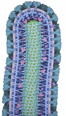
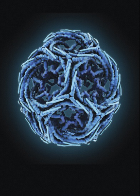
Two RCSB PDB images have been highlighted with 2016 Wellcome Image Awards. David S. Goodsell's painting of Ebola Virus has been selected as the overall winner, with Maria Voigt's molecular animation of Clathrin also recognized with an award.

Goodsell's painting was created for his RCSB PDB Molecule of the Month article on Ebola Virus, and also turned into a related video. This intricate painting shows this tiny, notoriously lethal virus in minute detail, using the structural information available in the Protein Data Bank archive.

Voigt's Clathrin illustration is part of RCSB PDB's traveling Art of Science exhibit, which uses the context of a community art gallery to introduce new audiences to to the beauty of structural biology. This exhibit is currently on display at the FlexSchool in Fanwood, NJ.

These are two of the 20 winning images selected as the best in science image making from all those acquired by the Wellcome Images picture library in the past year. The awards were presented at a ceremony at the Science Museum in London on March 15, where the images are now on show to the public. Other award-winning images include pathways of nerve fiber in the brain, delicate golden scales on a Madagascan sunset moth and a digitally reconstructed skeleton showing atheroma, the "furring up" of arteries that supply blood to the brain which can cause stroke.

16 science centers, museums and galleries will be displaying the winning images in their own styles to spark imaginations everywhere. This year the images will also be appearing as far afield as the Africa Centre for Population Health in South Africa, the Polytechnic Museum in Moscow, Russia, and the Massachusetts Institute of Technology.

The images are available to view on the Wellcome Image Awards website along with the stories behind the images and their creators. The images already feature in Wellcome Images collections, where they can be accessed and used along with more than 40,000 other contemporary biomedical and clinical images. The Awards were established in 1997 to reward contributors to the collection for their outstanding work.

Ebola Virus, by David S. Goodsell. The virus is surrounded by a membrane (pink/purple) stolen from an infected cell. This is studded with proteins from the virus (turquoise) which extend outwards and look like trees rooted in the membrane. These proteins attach to the cells that the virus infects. A layer of proteins (blue) supports the membrane on the inside. Genetic information (RNA; yellow) is stored in a cylinder (nucleocapsid; green) in the centre of the virus. More at Molecule of the Month on Ebola Virus Proteins.

Clathrin, by Maria Voigt. Clathrin is a protein found in cells which forms basket- or cage-like structures around small membrane sacs. These clathrin cages help bring molecules into the cell and then carry them around inside from one place to another. They also help sort through the molecules (which can include receptors and nutrients) so that different cargo is efficiently delivered to different destinations. Some disease-causing germs and toxins hijack this process and use it to infect cells. More at Molecule of the Month on Clathrin.


Past news and events have been reported at the RCSB PDB website and past Newsletters.