Annual Report Published
07/18
Download the 2016 Annual Report (PDF) for an overview of data deposition, query, outreach, and education activities.
This review highlights many RCSB PDB accomplishments, including educational materials focused on Diabetes and powerful 3D visualization tools.
wwPDB efforts, including deposition statistics and the OneDep system for deposition, validation, and biocuration, are also highlighted.
These bulletins provide a yearly snapshot of RCSB PDB activities and the state of the PDB archive. This edition is available as a PDF. If you would like a printed copy, please send your postal address to info@rcsb.org.
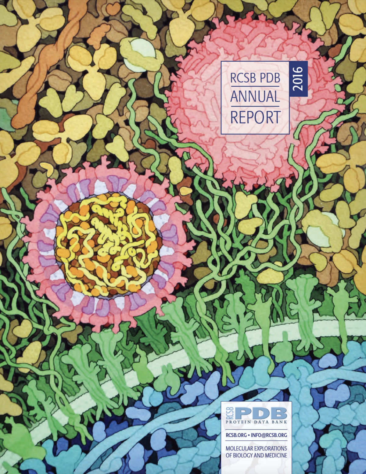 Zika virus is shown in cross section at center left. Visible on the periphery are envelope proteins (pink) and membrane proteins (magenta) embedded in a lipid membrane (light purple). Within the interior of the virus, the RNA genome (yellow) is associated with capsid proteins (orange). Two viruses are shown interacting with cell surface receptors (green) and are surrounded by blood plasma proteins outside the cell. This painting was recognized by the National Science Foundation (NSF) and Popular Science as one of the best science images of the year and selected as the “People’s Choice” in the illustration category for the 2017 Vizzies.
Past news and events have been reported at the RCSB PDB website and past Newsletters.
