News 2019
Season's Greetings
12/24
Play with Molecular Animations
12/17
Structural Biology and Nobel Prizes
12/09
Molecular Origami: Build a 3D Model of GPCR
12/03
Education Corner: Gaming Structural Biology for General Audiences (Part 2)
11/26
Introducing Mol*
11/19
New Papers on Molecular Visualization
11/12
Education Corner: Gaming Structural Biology for General Audiences (Part 1)
10/29
PDB Turns 48
10/20
Happy Birthday, Irving Geis
10/18
From the Bench to Molecule of the Month
10/15
Fall Newsletter Published
10/08
Structural Biology Pipeline Meets the Classroom: First Structure Released
09/25
Illustrate PDB Structures
09/17
Contact Customer Services with Questions and Feedback
09/10
Head Back to School with PDB-101
09/03
Poster Prize Awarded at ISMB
08/27
Poster Prize Awarded at ACA
08/20
Join Our Biocuration Team
08/06
Education Corner: How Does Life Work?
07/23
Beginner’s Guide to PDB Structures and the PDBx/mmCIF Format
07/16
Summer Newsletter Published
07/09
Create a Hemoglobin Bean Bag Toss
07/02
Learn About HIV and AIDS at PDB-101
06/25
New Flyer: Antibiotics in Action
06/04
Annual Report Published
05/21
Award-Winning Videos on Antibiotic Resistance
05/14
Education Corner: Exploring the Molecules of Biological Warfare in Virtual Reality
05/07
Vote Now for the Viewer's Choice Award
04/30
Celebrate DNA Day on April 25
04/23
Spring Newsletter Published
04/16
Take the Molecule of the Month User Survey and Enter to Win
04/09
High School Students: Submit Antibiotic Resistance Videos Before April 23
04/02
Molecular Landscapes and the Art of Science
03/26
The PDB Archive Reaches a Significant Milestone
03/19
New Video: Penicillin and Antibiotic Resistance
03/05
Superbugs! How Bacteria Evolve Resistance to Antibiotics
02/26
Join Our Team as a Biocurator
02/12
New Online Curriculum: The PDB Pipeline & Data Archiving
02/05
Education Corner: Improving Visual Literacy
01/29
Winter Newsletter Published
01/15
2018 FASEB BioArt Winner
01/08
2019: What is a protein?
01/01

Molecular Origami: Build a 3D Model of GPCR

12/03 

GPCRs are a large family of membrane-embedded receptors, with structural features that have been preserved through the course of evolution. This model represents the shared structural features of all GPCRs. With the extracellular N-terminus, the protein chain folds to form a bundle of seven transmembrane alpha helices connected by 3 intracellular and 3 extracellular loops with the C-terminus reaching inside the cell.

On the extracellular side, the helices form a cavity where ligands (e.g. endorphins, morphine, serotonin) bind. On the intracellular side, the receptor is coupled to G protein. When the receptor is activated by a ligand, the G protein splits in two parts which then activate other proteins in the internal signal transduction pathways.

Examples of GPCRs are opioid receptors, rhodopsin, or adrenergic receptors.

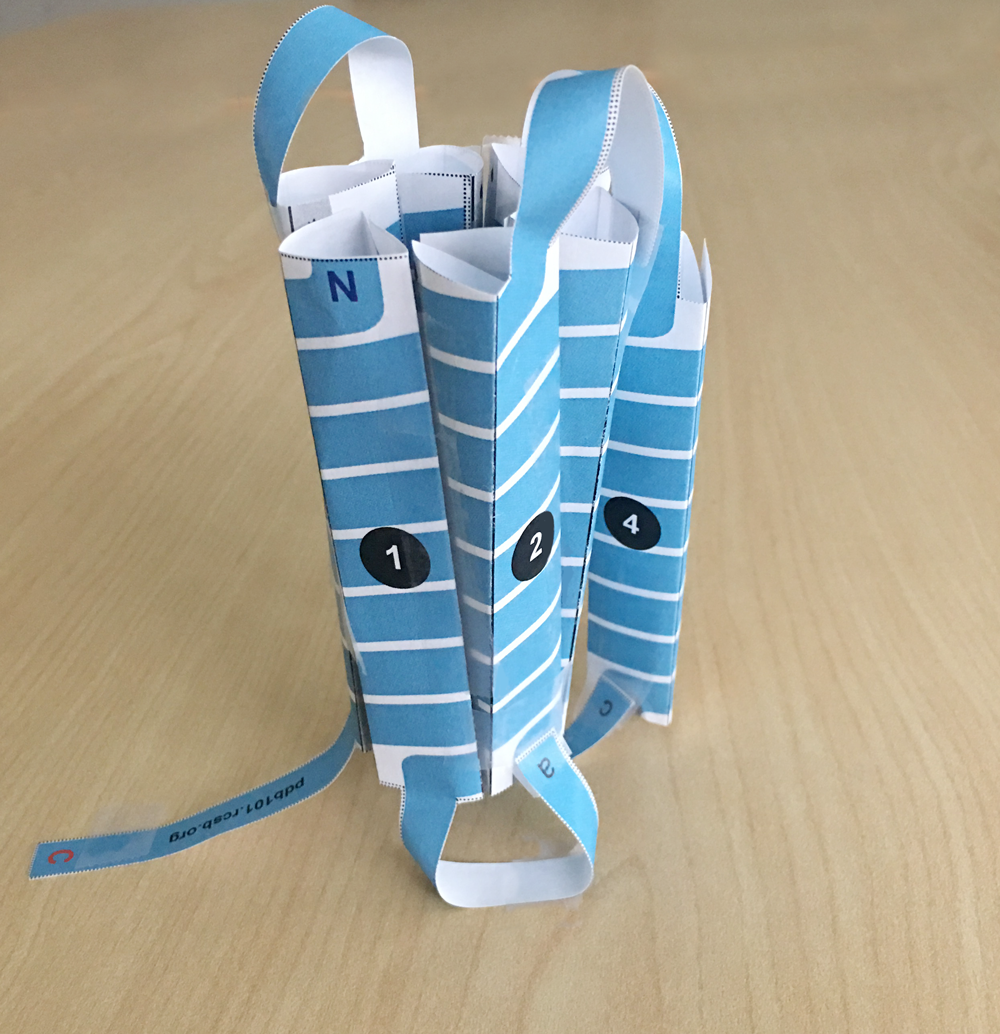
To build the GPCR paper model, download and print the template PDF. An instructional video is available.

Use the PDB-101 Browser to explore more resources and articles related to Drugs and the Brain.


Past news and events have been reported at the RCSB PDB website and past Newsletters.