Guide to Understanding PDB Data: Hierarchical Structure
12/15
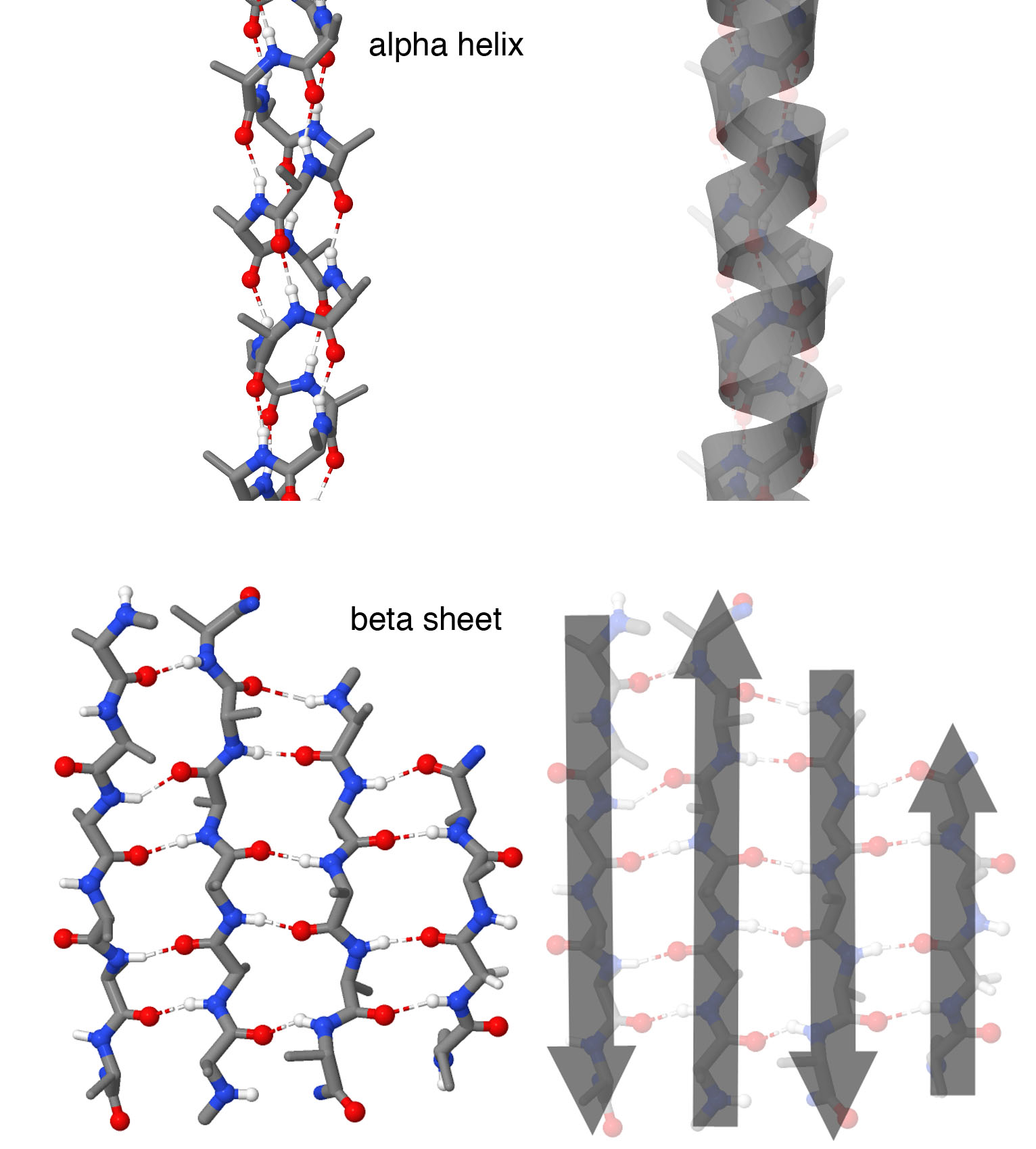 Atomic representations are shown at left, and cartoon representations are shown at left. Alpha helices form spring-shaped structures with each amino acid forming a hydrogen bond with an amino acid further down the chain. Beta strands are extended regions of the chain that form hydrogen bonds with neighboring beta strands, forming a beta sheet. In this structure, four beta strands form the sheet with neighboring strands running in opposite directions, as shown by the cartoon arrows. Note that in these illustrations, side chains are not shown on each amino acid, to make it easier to see the backbone structure. Structures taken from PDB ID 1ic2 and 4gcr.
The constantly-growing PDB is a reflection of the research that is happening in laboratories across the world. This can make it both exciting and challenging to use the database in research and education.
PDB-101's Guide to Understanding PDB Data was created to help users navigate through the contents of the archive without having a detailed background in structural biology.
Topics cover biological assemblies, molecular graphics programs, R-value and R-free, and more.
A new chapter highlights the Hierarchical Structure of Proteins. Topics in this article include:
- Why is Understanding Protein Folding Important?
- Hierarchical Structure
- Visualization of Hierarchical Structure
- Classifying Protein Folds
Past news and events have been reported at the RCSB PDB website and past Newsletters.
