GLP-1 Receptor
Glucagon-like peptide 1 receptor (GLP-1R) is a G-Protein coupled receptor found on the cell membranes of pancreatic and other cells. It is the target for several FDA-approved antidiabetic medications called incretin mimetics:
| a. Abliglutide (Tanzeum) | c. Liraglutide (Victoza) | e. Exenatide (Byetta, Bydureon) | g. Tirzepatide (Mounjaro) |
| b. Dulaglutide (Trulicity) | d. Lixisenatide (Adlyxin) | f. Semaglutide (Ozempic, Rybelsus, Wegovy) |
These incretin mimetics are glucagon-like peptide 1 (GLP-1) analogs that mimic the physiological action of GLP-1 and thereby potentiates the incretin effect in type 2 diabetic patients. These agents work in the same pathway as DPP-4 inhibitors but are generally considered more potent.
Function
The GLP-1 receptor (GLP-1R) is a seven-segmented transmembrane protein with an extracellular amino terminus and an intracellular carboxyl terminus. It binds to the glucagon-like peptide 1 (GLP-1) hormone and potentiates the synthesis and secretion of insulin from pancreatic β-cells in a glucose-induced manner (Underwood et al., 2009). GLP-1R plays a critical role in the signaling cascades leading to insulin release in response to an increase in blood glucose levels.
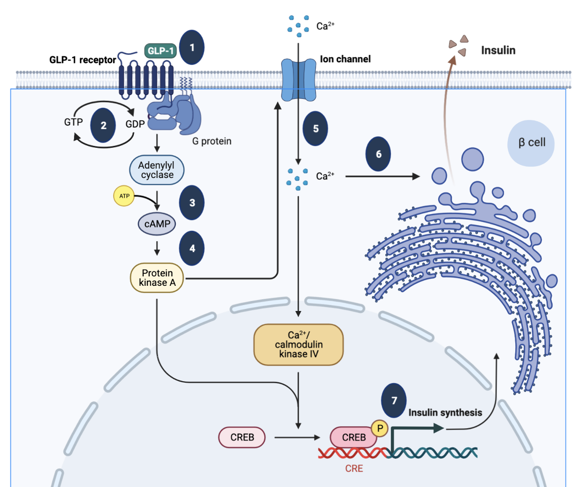
GLP-1 receptor belongs to class B1 of the G-protein-coupled receptors (GPCR), a subfamily characterized by not only a large N-terminal extracellular ligand binding domain and seven-transmembrane topology, but also a cAMP-mediated intracellular signaling pathway. The GLP-1R interacts with structurally related peptide hormone ligands, such as glucagon-like peptide-1 (GLP-1), glucose-dependent insulinotropic polypeptide (GIP), parathyroid hormone, etc. (Runge et al., 2008). Key steps in GLP-1 signaling, as shown in Figure 1, include:
- The ligand GLP-1 binds to the GLP-1R, a G-coupled protein receptor.
- The GLP-1R interacts with the heterotrimeric G-protein composed of α, β and γ subunits. A guanosine diphosphate (GDP) is bound to the α-subunit. When the ligand binds to the extracellular domain of the receptor, the G-protein undergoes conformational change and GTP replaces the GDP on the α-subunit.
- The GTP-bound α-subunit dissociates from the trimer and activates adenylyl cyclase, which in turn produces large amounts of cyclic AMP (cAMP) from ATP within the cell.
- The cAMP then binds to and activates target proteins such as Protein Kinase A, which phosphorylates specific cells in the cell via a signaling cascade pathway.
- Activation of PKA facilitates membrane depolarization, which consequently opens voltage‐gated Ca2+ channels, allowing an influx of Ca2+ ions.
- Increased intracellular Ca2+ concentrations trigger fusion of insulin-containing granules with the plasma membrane and insulin secretion from the β cells.
- Increased Ca2+ levels also promote transcription of the proinsulin gene, thereby increasing the insulin content of the β cell.
|
| Figure 1. Molecular mechanisms underlying the insulinotropic effects of glucagon-like peptide-1. Figure inspired by Mayendraraj et al., 2022 and drawn using Biorenderer. |
Structure
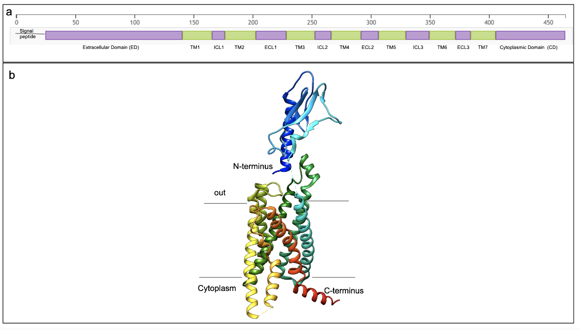
The GLP-1R protein has a signal peptide (amino acids 1-23) that is cleaved as the rest of the protein (amino acids 24-463) is targeted to the endoplasmic reticulum to form a seven transmembrane helix containing protein (see extracellular (E), cytoplasmic (C), and membrane proteins (M) marked in Figure 2a). The EM structures (Figure 2b) of the receptor revealed that it is composed of:
- The extracellular domain (E) of the GLP-1 receptor binds the GLP-1 protein
- A seven transmembrane helical domain (TM1-7) with the ɑ-helices separated by three intracellular loops (IC1-3) and three extracellular loops (EC1-3)
- An intracellular (Cytoplasmic) C-terminus that interacts with G-Protein which is responsible for signaling
|
| Figure 2: GLP-1R architecture: a. Linear schematic of the GLP-1 receptor showing signal peptide, extracellular domain (E), transmembrane helices (TM 1-7), extracellular loops (ECL1-3), intracellular loops (ICL1-3), and cytoplasmic domain (C) . b. Ribbon representation of the extracellular domains of GLP-1 receptor protein (PDB ID 6x18, Zhang et al., 2020). The structure is colored using the rainbow color scheme with the N-terminus (extracellular) is colored blue and the C-terminus (cytoplasmic) is colored in red. Note: only the receptor protein chain is shown here. The GLP-1 protein, bound to the extracellular domain, and G-protein related protein chains bound to the cytoplasmic domain are hidden for clarity. |
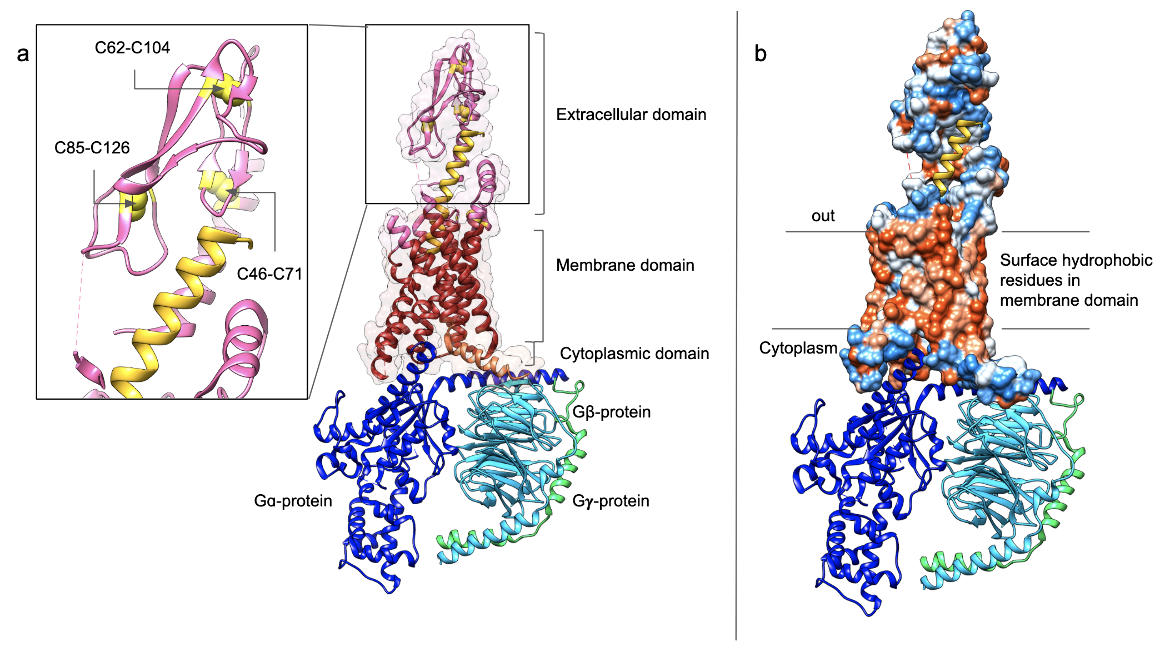
The extracellular domain: The N-terminal ~120 amino acids of the extracellular domain of the GLP-1R protein has one long and one short helix, two beta strands forming a sheet and several loops joining these secondary structural elements. Three short loops between the transmembrane helices of this receptor also make up the extracellular domain. This domain is stabilized by three disulphide bonds between amino acid residues 46-71; 62-104; and 85-126 (see Figure 3a inset). In addition, there are a few conserved residues (e.g., Asp67, Trp72, Pro86, Gly108, and Trp110) that form specific interactions within the extracellular domain (see Figure 4 insets).
The transmembrane helices: Like any other G-protein coupled receptor, this protein has seven transmembrane helices (TM1-7) with surface exposed hydrophobic amino acids in the membrane region (see Figure 3b).
The cytoplasmic domain: This region of the receptor interacts with the G-proteins. Binding of the ligand (GLP-1) or its analog on the extracellular side of the protein induces conformational changes that are sensed by the G-proteins on the cytoplasmic face - initiating a signaling cascade (see Figure 3 a and b).
|
| Figure 3: 3D structure of GLP-1R bound to GLP-1 (PDB ID 6x18, Zhang et al., 2020) a. Ribbon and surface outline representations of the receptor showing extracellular region in pink, transmembrane region in red, and cytoplasmic regions of the receptor in orange. The G-proteins (ɑ, β, and 𝛄) are shown in dark blue, light blue, and green, respectively. The bound GLP-1 peptide is colored yellow. The inset shows the 3 disulfide bridges in the extracellular domain. b. The surface residues are shown colored according to the hydrophobicity scale of Kyte and Doolittle - where orange indicates hydrophobic and blue indicates hydrophilic. The G-proteins are colored using the same color scheme as in figure a. |
The primary function of the GLP-1R is to bind the GLP-1 protein and its analogs and initiate signaling inside pancreatic β-cells to release insulin. Although this receptor can bind a variety of different peptide and small molecule ligands, here we describe the function of GLP-1R function as – a. GLP-1 binding (outside the cell), and b. G-protein signaling (inside the cell).
GLP-1 Binding
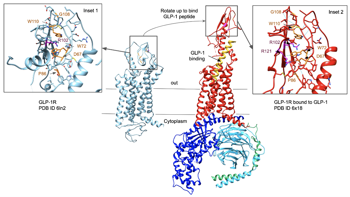
The EM structure of the GLP-1 apo receptor (i.e., receptor in the absence of any ligand), shows that the N-terminal domain adopts a closed or inactive conformation, where a cap-like structure blocks access to the peptide binding ( PDB ID 6ln2, Wu et al., 2020, Figure 4a). The cap-like structure includes some of the conserved amino acids forming pi-cation interactions between Trp72, Arg102, and Trp110 (Figure 4 insets 1 and 2).
|
| Figure 4: Comparison of the GLP-1 receptor alone vs the receptor bound to the GLP-1 peptide to the extracellular domain and G-proteins bound to the cytoplasmic domain. The Apo-receptor (PDB ID 6ln2, Wu et al., 2020) is colored in blue, while the GLP-1 receptor bound to the peptide (PDB ID 6x18, Zhang et al., 2020) is colored red. The G-proteins and the peptide bound to the GLP-1 receptor are colored using the same color code as in Figure 3. |
Peptide binding is facilitated by a major conformational change, where the N-terminal domain opens up to bind the GLP-1 peptide (Figure 4). These conformational changes are stabilized by additional noncovalent interactions involving the conserved residues, such as the Arg121 making hydrogen bonds with Asp67 to further strengthen the pi-cation interactions involving Trp72, Arg102, and Trp110 (see Figure 4 inset 2).
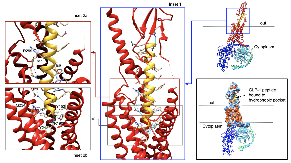
Although studies determining the structure of GLP-1 bound to the N-terminal, extracellular domain of the receptor showed that the residues 13-33 of the GLP-1 peptide analogs form a helical structure (PDB ID 3iol, Underwood et al., 2010), recent EM structures of the whole receptor bound to the GLP-1 protein (PDB ID 6x18, Zhang et al., 2020) shows that the entire GLP-1 peptide forms a long helix as it binds to its receptor. The GLP-1 peptide interacts with the receptor via several polar and hydrophobic interactions (see Figure 5). Examples of the non-polar interactions between side chains atoms of GLP-1 and the receptor protein include His7 of GLP-1 forming a hydrogen bond with Gln234 of the receptor; Glu9 of the GLP-1 forms a direct hydrogen bond with Tyr152, an ionic interactions with Arg190, and a water mediated hydrogen bond with Tyr241; Ser17 of GLP-1 forming hydrogen bonds with Arg299 and Tyr205; and Glu21 of GLP-1 forming ionic interactions with Arg299. Alanine mutation studies have shown that removing these interactions does impact GLP-1 receptor binding.
|
| Figure 5: Interactions stabilizing the binding of GLP-1 (yellow helix) to its receptor (red colored protein) (PDB ID 6x18, Zhang et al., 2020). Ribbon representation of the overall structure including the G-protein chains are shown in the top right corner. Expansions of boxed regions are shown in the insets 1, 2a and 2b. The bottom right figure shows the hydrophobic surface in the peptide binding pocket. The G-protein chains are shown using the same color scheme as in Figure 3. |
GPCR Activation
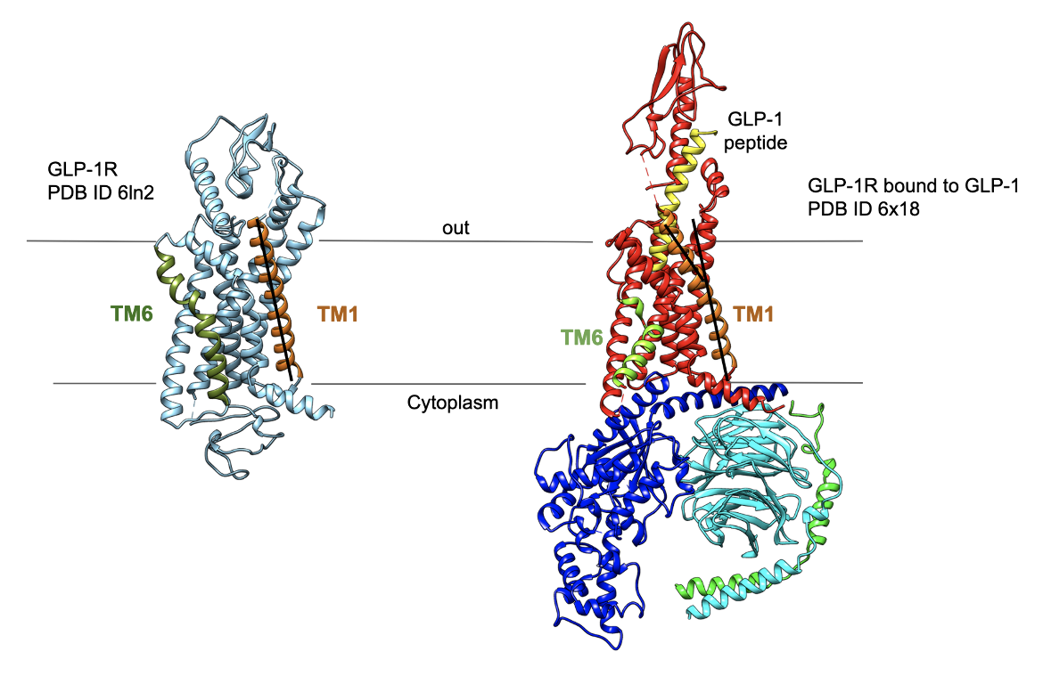
The conformational changes in the receptor associated with GLP-1 binding to its extracellular face is communicated to the cytoplasmic face by the transmembrane helices rotating relative to each other (Zhang et al., 2020). For example, after GLP-1 binding the transmembrane helices 1 (TM1) rotates clockwise when looking down the membrane helices from the extracellular face (see TM1 marked in Figure 6) and a kink is introduced in the transmembrane helix 6 (TM6), as is typical for all class B1 GPCRs. These conformational changes impact the binding of the Gɑ-protein through a series of hydrophobic interactions and some specific non-polar interactions. In turn, the G-proteins activate adenylyl cyclase, leading to a signal transduction. Lear more about G-protein signaling.
|
| Figure 6: Comparison of the GLP-1R structures without and with GLP-1 bound. The ribbon representation of the apo receptor shows the transmembrane helices TM1 and TM6 relatively straight (Wu et al., 2020), while that of the GLP-1 peptide bound form shows twists (in TM1) and a kink (in TM6) (Zhang et al., 2020). The protein chains are colored using the same color scheme as in Figure 4. |
Pharmacological Implications
In patients with diabetes, the incretin effect is reduced. DPP-4 inhibitors inhibit the degradation of native incretins, i.e. GIP and GLP-1, to extend the half-life of these hormones and thereby pharmacologically enhance their physiological effects. Another way to approach this outcome would be to identify agonists that act on the GLP-1 receptor to produce the same pharmacological outcome. Elucidation of the structure of GLP-1R to understand its interaction with GLP-1 and its peptidic and small molecule agonists have made this a promising approach for treating type 2 diabetes. Today several incretin mimetics are in clinical use for stimulating insulin synthesis and secretion.
Other Considerations
While action of GLP-1 on pancreatic β-cells increases insulin release, this hormone and its analogs have key impact on gastric emptying and satiety. In recent years the incretin mimetics have also been used in weight management. Note that GLP-1 is not the only hormone that binds to and signals via the GLP-1R. A variety of other natural peptides have also been shown to bind this receptor. While the gastric emptying function of incretin mimetics may facilitate weight loss, it may have gastrointestinal effects and also impact the action of other medications taken orally.
References
Mayendraraj, A., Rosenkilde, M.M., Gasbjerg, L.S. (2022) GLP-1 and GIP receptor signaling in beta cells - A review of receptor interactions and co-stimulation. Peptides. 151, 170749. https://doi.org/10.1016/j.peptides.2022.170749
Runge, S., Thøgersen, H., Madsen, K., Lau, J., Rudolph, R. (2008) Crystal structure of the ligand-bound glucagon-like peptide-1 receptor extracellular domain. J Biol Chem., 283, 11340-7. https://doi.org/10.1074/jbc.m708740200
Underwood, C.R., Garibay, P., Knudsen, L.B., Hastrup, S., Peters, G.H., Rudolph, R., Reedtz-Runge, S. (2010) Crystal structure of glucagon-like peptide-1 in complex with the extracellular domain of the glucagon-like peptide-1 receptor. J Biol Chem. 285, 723-30. https://doi.org/10.1074/jbc.m109.033829
Wu, F., Yang, L., Hang, K., Laursen, M., Wu, L., Han, G.W., Ren, Q., Roed, N.K., Lin, G., Hanson, M.A., Jiang, H., Wang, M.W., Reedtz-Runge, S., Song, G., Stevens, R.C. (2020) Full-length human GLP-1 receptor structure without orthosteric ligands. Nat Commun. 11, 1272. https://doi.org/10.1038/s41467-020-14934-5
Zhang, X., Belousoff, M.J., Zhao, P., Kooistra, A.J., Truong, T.T., Ang, S.Y., Underwood, C.R., Egebjerg, T., Šenel, P., Stewart, G.D., Liang, Y.L., Glukhova, A., Venugopal, H., Christopoulos, A., Furness, S.G.B., Miller, L.J., Reedtz-Runge. S., Langmead, C.J., Gloriam, D.E., Danev, R., Sexton, P.M., Wootten, D. (2020) Differential GLP-1R Binding and Activation by Peptide and Non-peptide Agonists. Mol Cell. 80, 485-500. https://doi.org/10.1016/j.molcel.2020.09.020
August 2023 Jennifer Jiang and Dr. Shuchismita Dutta; Reviewed by Dr. Joseph D. Ho
http://dx.doi.org/10.2210/rcsb_pdb/GH/DM/drugs/in/GLP-1R
