Nobel Prizes and PDB structures
award-winning research
Many Nobel Prize awards in physics, chemistry, and physiology/medicine have recognized scientific achievements that can be associated with structural biology. The articles and resources below highlight many of these PDB structures and related experimental techniques.
Molecule of the Month Articles (31)
 |
Adenylyl Cyclase
Adenylyl cyclase creates second messengers to amplify signals from G-protein coupled receptors |
 |
Adrenergic Receptors
Adrenaline stimulates a G-protein-coupled receptor, priming us for action |
 |
Aminopeptidase 1 and Autophagy
Aminopeptidase 1 is delivered inside the cell using the machinery of autophagy |
 |
Antibodies
Antibodies search for foreign molecules in the blood |
 |
Aquaporin
Aquaporins create a channel for water molecules to cross through cell membranes |
 |
ATP Synthase
ATP synthase links two rotary motors to generate ATP |
 |
Bacteriorhodopsin
Bacteriorhodopsin pumps protons powered by green sunlight |
 |
Capsaicin Receptor TRPV1
TRPV1 is an ion channel that senses heat and contributes to pain sensation. |
 |
Cascade and CRISPR
Cascade and CRISPR help bacteria remember how to fight viral infection |
 |
Circadian Clock Proteins
Circadian clock proteins measure time in our cells |
 |
Click Chemistry
A modular approach to chemistry simplifies the construction of complex protein-targeting molecules. |
 |
Directed Evolution of Enzymes
Biological evolution is being harnessed in the lab to create new enzymes. |
 |
DNA
Atomic structures reveal how the iconic double helix encodes genomic information |
 |
FOXP3
A master transcriptional regulator of immune tolerance |
 |
G Proteins
G proteins receive signals from cellular receptors and deliver them inside the cell |
 |
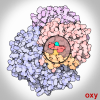
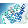
Hemoglobin
Hemoglobin uses a change in shape to increase the efficiency of oxygen transport |
 |
Hepatitis C Virus Protease/Helicase
Structures of hepatitis C viral proteins have led to the discovery of direct-acting antivirals. |
 |
Hypoxia-Inducible Factors
HIF-α is a molecular switch that responds to changing oxygen levels. |
 |
Myoglobin
Myoglobin was the first protein to have its atomic structure determined, revealing how it stores oxygen in muscle cells. |
 |
PDB Pioneers
A dozen historic structures set the foundation for the PDB archive |
 |
Photosystem I
Photosystem I captures the energy in sunlight |
 |
Photosystem II
Photosystem II captures the energy from sunlight and uses it to extract electrons from water molecules |
 |
Piezo1 Mechanosensitive Channel
Mechanosensitive ion channels give our cells a sense of touch. |
 |
Potassium Channels
Potassium channels allow potassium ions to pass, but block smaller sodium ions |
 |
Ribonuclease A
Ribonuclease cuts and controls RNA |
 |
Ribosomal Subunits
Atomic structures of the ribosomal subunits reveal a central role for RNA in protein synthesis |
 |
Ribosome
Ribosomes are complex molecular machines that build proteins |
 |
RNA Polymerase
RNA polymerase transcribes genetic information from DNA into RNA |
 |
Spliceosomes
Cryoelectron microscropy is revealing how spliceosomes cut-and-paste messenger RNA molecules. |
 |
Telomerase
Telomerase maintains the ends of our chromosomes. |
 |
Tobacco Mosaic Virus
A cylindrical arrangement of proteins protects a long strand of RNA in TMV |
Learning Resources (25)
 |
DNA
Paper Model
Atomic structures reveal how the iconic double helix encodes genomic information
|
 |
Green and Red Fluorescent Proteins
Paper Model
A tiny fluorescent protein from jellyfish has revolutionized cell biology
|
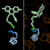
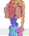
| G Protein-Coupled Receptor (GPCR)
Paper Model
GPCRs are a large family of membrane-embedded receptors, with structural features that have been preserved through the course of evolution. This model represents the shared structural features of all GPCRs. With the extracellular N-terminus, the protein chain folds to form a bundle of seven transmembrane alpha helices connected by 3 intracellular and 3 extracellular loops with the C-terminus reaching inside the cell.
|
|
 |
Expanding Boundaries of Complexity with 3DEM
Flyer
3D electron microscopy (3DEM) is revolutionizing the field of structural biology.
|
 |
ADN: El Acido Desoxirribonucleico (Spanish)
Flyer
Atomic structures reveal how the iconic double helix encodes genomic information
|
| 2021 Calendar Celebrating 50 Years of Protein Data Bank
Calendar
2021 Calendar
|
|
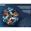 |
Insulin and Dorothy Hodgkin
Flyer
|
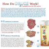 |
How do Drugs Work?
Flyer
PDB structures are used to discuss antibiotics and antivirals, chemotherapy, drug metabolism, drugs of signaling proteins, and lifestyle drugs.
|
 |
G-Protein Coupled Receptors
Flyer
In honor of the 2012 Nobel Prize in Chemistry.
|
| Computed Structure Models and PDB Experimental Structures Poster
Poster
Simultaneous delivery of PDB data and Computed Structure Models on RCSB.org provides access to 3D structural information from across the human proteome, model organisms, and selected pathogens
|
|
 |
Toll-like Receptors
Flyer
In honor of the 2011 Nobel Prize in Physiology or Medicine.
|
 |
The Ribosome
Flyer
This flyer commemorates the 2009 Nobel Prize in Chemistry for studies of the structure and function of the ribosome.
|
 |
How Do Drugs Work?
Poster
PDB structures are used to discuss antibiotics and antivirals, chemotherapy, drug metabolism, drugs of signaling proteins, and lifestyle drugs.
|
 |
ATP synthase
GIF
ATP synthase
|
 |
Oxygen Binding in Hemoglobin
GIF
Hemoglobin uses a change in shape to increase the efficiency of oxygen transport.
|
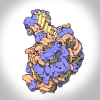 |
Ribosomal Subunits
GIF
Atomic structures of the ribosomal subunits reveal a central role for RNA in protein synthesis. Ribosomes are complex molecular machines that build proteins.
|
 |
Celebrating 50 Years of the Protein Data Bank Archive
Video
In 1971, the structural biology community established the single worldwide archive for macromolecular structure data–the Protein Data Bank (PDB). From its inception, the PDB has embraced a culture of open access, leading to its widespread use by the research community. PDB data are used by hundreds of data resources and millions of users exploring fundamental biology, energy, and biomedicine.
This video looks at the history and the milestones that shaped the PDB into the leading resource for research and education it is today.
|
 |
Methods for Determining Structure
Guide
|
 |
3D Print: Hemoglobin
Other Resource
Download curated file of hemoglobin structure for 3D printing.
|
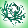 |
Exploring Green Fluorescent Protein Tutorial
Other Resource
Green Fluorescent Protein (GFP) is a well-studied protein that exhibits fluorescence when exposed to ultraviolet or blue light, making it a powerful tool in molecular biology and biotechnology. Learn how to explore GFP using tools from the RCSB PDB with this tutorial.
|
| Commemorating 75 Years of Discovery and Innovation at the NSF
Other Resource
Download images celebrating NSF and PDB milestones
|
|
| Molecular Backgrounds For Virtual Meetings
Other Resources
Download images created by David Goodsell to add a molecular backdrop to your next virtual meeting. Click on the image to expand.
|
|
 |
Structural Biology and Nobel Prizes
Other Resource
Many Nobel Prizes have recognized achievements made in molecular biology, structural biology, and related research.
|
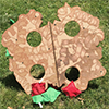 |
Hemoglobin Bean Bag Toss
Other Resource
|
| Exploring Structural Biology with Computed Structure Models (CSMs)
Article
Computational methods like AlphaFold2 and RoseTTAFold2 use structures in the PDB archive to predict the folding of proteins.
|
Geis Digital Archive (5)
 |
Myoglobin
Geis highlights the hundreds of chemical bonds in the lattice of myoglobin. |
 |
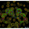
Ribonuclease S
Geis illustrates the structure of the ribonuclease S that highlights the dinucleotide RNA substrate in red and the four disulfide bonds in yellow. |
 |
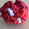
Hemoglobin
Geis illustrates the hemoglobin molecule as four symmetrically arranged myoglobins. Since it is responsible to the transport of oxygen, it can change from an oxygen-binding configuration to an oxygen-releasing configuration in response to the demand for oxygen. |
 |
B-DNA
Geis illustrates B-DNA in blue looking from above, through the double helix. The two bases on top are highlighted in white to distinguish one individual section of the layered scene. |
 |
DNA
Geis illustrates a double helix in his depiction of DNA. He portrays the helices with a soft ribbon structure. The white "box-like" structures represent a base pair in the DNA strand.
|
