Biomolecules
the building blocks of life
Most of the molecular machines built by cells are composed of protein and nucleic acid chains. Atomic structures have revealed how these chains are built and how they fold into functional molecular machines.
Molecule of the Month Articles (14)
 |
Aconitase and Iron Regulatory Protein 1
Aconitase performs a reaction in the citric acid cycle, and moonlights as a regulatory protein |
 |
Adenine Riboswitch in Action
XFEL serial crystallography reveals what happens when adenine binds to a riboswitch |
 |
Aspartate Transcarbamoylase
Key biosynthetic enzymes are regulated by their ultimate products through allosteric motions. |
 |
Circadian Clock Proteins
Circadian clock proteins measure time in our cells |
 |
Collagen
Sturdy fibers of collagen give structure to our bodies |
 |
Concanavalin A and Circular Permutation
For some proteins, clipped and reassembled sequences can produce the same 3D shape |
 |
Crystallins
A concentrated solution of crystallins refracts light in our eye lens |
 |
Designed Proteins and Citizen Science
What if people with no formal experience in science could help to improve or even rewrite nature, simply by playing a game? |
 |
DNA
Atomic structures reveal how the iconic double helix encodes genomic information |
 |
Fifty Years of Open Access to PDB Structures
The Protein Data Bank is celebrating its golden anniversary! |
 |
Globin Evolution
The mechanisms of molecular evolution are revealed in globin sequences and structures. |
 |
Hemoglobin
Hemoglobin uses a change in shape to increase the efficiency of oxygen transport |
 |
Insulin
The hormone insulin helps control the level of glucose in the blood |
 |
Quasisymmetry in Icosahedral Viruses
Viruses use quasisymmetry to build large capsids out of many small subunits |
Learning Resources (37)
 |
Creating Paper Models of Protein Domains
Paper Model
Download printable alpha helices and beta strand templates for hands-on exploration of many protein folding domains.
|
 |
DNA
Paper Model
Atomic structures reveal how the iconic double helix encodes genomic information
|
| Dengue Virus
Paper Model
Atomic structures of dengue virus are giving new hope for creation of a vaccine
|
|
 |
HIV Capsid
Paper Model
At the center of HIV, an unusual cone-shaped capsid protects the viral genome and delivers it into infected cells
|
 |
Insulin
Paper Model
Learn about insulin, a peptide hormone that plays a critical role in our ability to use glucose from the food that we eat
|
| tRNA
Paper Model
Transfer RNA translates the language of the genome into the language of proteins
|
|
 |
ADN: El Acido Desoxirribonucleico (Spanish)
Flyer
Atomic structures reveal how the iconic double helix encodes genomic information
|
 |
2022 Calendar: Revolutionizing structural biology with 3DEM
Calendar
This calendar features twelve molecular assemblies studied by 3DEM. They further our understanding of biology and serve as a gateway to advances in technology and medicine.
|
| 2021 Calendar Celebrating 50 Years of Protein Data Bank
Calendar
2021 Calendar
|
|
 |
Molecular Machinery: A Tour of the Protein Data Bank
Flyer
The enormous range of molecular shapes and sizes in the PDB archive is illustrated using 96 structures, depicted relative to the cellular membrane.
|
| 2020 Calendar: 20 Years of Molecule of the Month
Calendar
This calendar celebrates the RCSB PDB Molecule of the Month series that has introduced millions of visitors to the shape and function of the 3D structures archived in the PDB since its launch in January 2000.
|
|
| 2019 Calendar: What is a Protein?
Calendar
Learn about protein structure and function with this 2019 calendar and related materials.
|
|
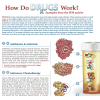 |
How do Drugs Work?
Flyer
PDB structures are used to discuss antibiotics and antivirals, chemotherapy, drug metabolism, drugs of signaling proteins, and lifestyle drugs.
|
 |
What is a Protein?
Flyer
Learn about protein structure and function with this overview.
|
 |
Virus Structures
Flyer
Learn about polyhedral, helical, complex, and enveloped viruses with examples drawn at approximately 900,000x magnification. Created in collaboration with EMDataBank.
|
 |
The Structures of the Citric Acid Cycle
Flyer
Also known as the Krebs cycle or the tricarboxylic acid cycle, the citric acid cycle is at the center of cellular metabolism. Learn about the structures involved in this metabolic pathway.
|
 |
Folding of Protein Domains
Poster
This poster highlights common arrangements of secondary structure elements in protein domains as defined using the CATH classification scheme.
|
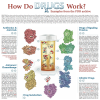 |
How Do Drugs Work?
Poster
PDB structures are used to discuss antibiotics and antivirals, chemotherapy, drug metabolism, drugs of signaling proteins, and lifestyle drugs.
|
 |
Molecular Machinery: A Tour of the Protein Data Bank
Poster
The enormous range of molecular shapes and sizes in the PDB archive is illustrated using 96 structures, depicted relative to the cellular membrane.
|
 |
The Structural Biology of HIV (Poster)
Poster
A painting of the HIV virus surrounded by the atomic structures from the PDB.
|
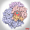 |
Oxygen Binding in Hemoglobin
GIF
Hemoglobin uses a change in shape to increase the efficiency of oxygen transport.
|
 |
What is a Protein? (Video)
Video
Proteins play countless roles throughout the biological world, from catalyzing chemical reactions to building the structures of all living things. Despite this wide range of functions all proteins are made out of the same twenty amino acids, but combined in different ways. The way these twenty amino acids are arranged dictates the folding of the protein into its primary, secondary, tertiary, and quaternary structure. Since protein function is based on the ability to recognize and bind to specific molecules, having the correct shape is critical for proteins to do their jobs correctly. Learn more about the relationship between protein structure and function in this video.
|
 |
Celebrating 50 Years of the Protein Data Bank Archive
Video
In 1971, the structural biology community established the single worldwide archive for macromolecular structure data–the Protein Data Bank (PDB). From its inception, the PDB has embraced a culture of open access, leading to its widespread use by the research community. PDB data are used by hundreds of data resources and millions of users exploring fundamental biology, energy, and biomedicine.
This video looks at the history and the milestones that shaped the PDB into the leading resource for research and education it is today.
|
 |
Interactive Tour of PDB's Molecular Machinery
Viewer
This interactive view of molecular machinery in the PDB archive lets users select a structure, access a 3D view of the entry using the NGL Viewer, read a brief summary of the molecule’s biological role, and access the corresponding PDB entry and Molecule of the Month article.
|
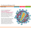 |
The Structural Biology of HIV
Interactive
PDB-101 highlights how these PDB structures have increased our understanding of HIV in an interactive animation and a poster.
|
 |
Coloring Molecular Machinery
Coloring Book
This is a molecular coloring book for all artists.
|
 |
Discovering Biology Through Crystallography
Coloring Book
Color the diverse 3D shapes studied by crystallographers. Created with support from the ACA. Available as a PDF and individual images.
|
| Protein Hierarchical Structure
Guide
Structures in the Protein Data Bank archive have revealed that folded proteins have several levels of hierarchical organization.
|
|
 |
Small Molecule Ligands
Guide
|
| Exploring Carbohydrates
Guide
The RCSB provides many tools to explore the functional roles of carbohydrates.
|
|
 |
Insulin Exercise
Curriculum
This exercise is a part of RCSB PDB Curricular Module Biomolecular Structures and Models and was developed as part of the RCSB Collaborative Curriculum Development Program 2015.
|
| Lesson Plans
Other Resource
|
|
| Molecular Backgrounds For Virtual Meetings
Other Resources
Download images created by David Goodsell to add a molecular backdrop to your next virtual meeting. Click on the image to expand.
|
|
| PDB50 the Game
Other Resource
A PDB “worker placement” board game that explores the process of structure discovery
|
|
| Structural Biology Playing Cards
Other Resources
Downloadable cards that highlight structures involved in infrastructure, energy, information, and catalysis
|
|
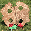 |
Hemoglobin Bean Bag Toss
Other Resource
|
| Exploring the Structural Biology of Evolution
Article
Evolution is a natural process that produces improved organisms without any need for intelligent intervention.
|
Curriculum Resources (13)
Structural Biology Highlights (6)
Geis Digital Archive (6)
 |
Deoxyhemoglobin
Geis illustrates deoxyhemoglobin in blue, a color associated with the absence of the oxygen molecule. Hemoglobin undergoes a conformational change in the absence of oxygen to produce this conformer. In the center is a red area showing the footprint of the space between the beta chains in the oxygenated state. The beta chains move apart in the deoxygenated state.
|
 |
DPG-Hemoglobin Complex
Geis illustrates the complex of DPG (2,3-diphosphoglycerate) with hemoglobin as a co-crystal. The charged amino acid residues stabilizing the complex with DPG are drawn in blue. |
 |
Oxyhemoglobin
Geis illustrates the hemoglobin molecule in its oxygenated state.
|
 |
Deoxyribonucleic Acid (DNA)
Geis illustrates three possible forms of deoxyribonucleic acid (DNA). He highlights the differences between each structure by displaying them in a side-by-side manner. |
 |
B-DNA
Geis illustrates B-DNA in blue looking from above, through the double helix. The two bases on top are highlighted in white to distinguish one individual section of the layered scene. |
 |
A-DNA
Geis uses a thin ball and stick representation of a section of A-DNA, the more compact conformation of DNA less often seen in biological systems. He draws it from a perspective looking down into the double helix, showing the increase in diameter of the middle of the helix from the B form DNA.
|


